An umbilical hernia in kittens and puppies happens prior to birth when there is a weakness or opening in the muscle wall of the abdomen where the umbilical blood vessels pass.
Frequently abdominal fat is in the hernia but the skin is intact across the hernia, so there are no exposed abdominal organs. The fat, called the omentum , may be part of the double layer of fatty tissue that covers and supports the organs and intestines in the lower abdomen. The abdominal fat could also be part of the falciform ligament which connects the liver to the abdomen. Rarely, intestines may slip into the hernia.
Umbilical hernias in puppies and kittens are relatively common and typically harmless, however, if this happens, you’ll want to educate yourself and contact your vet to make sure no further complications occur.
What Causes Umbilical Hernia in Puppies & Kittens?
It is generally considered that umbilical hernias have a genetic basis. Despite the wish that umbilical hernias are caused by trauma to the umbilical stump at birth, this is rarely the case. Veterinary staff is very careful to tie and handle umbilical cords carefully. Even when a female is aggressive while chewing the cords, they do not result in hernias.
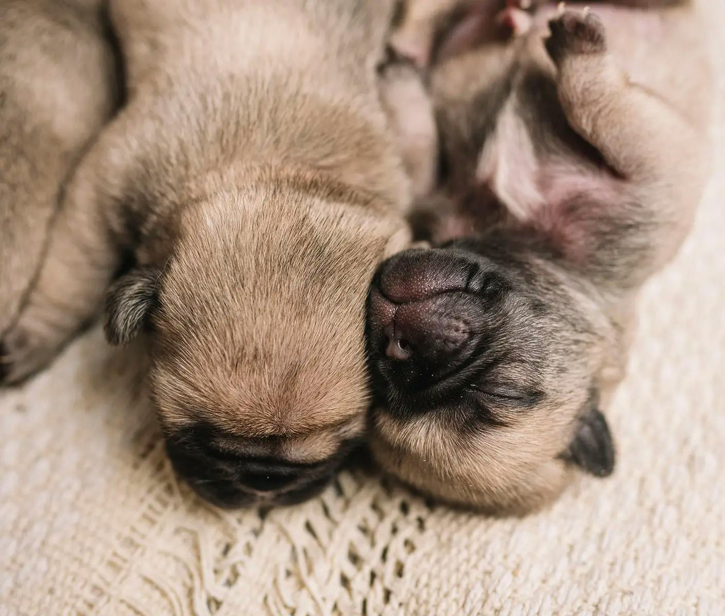
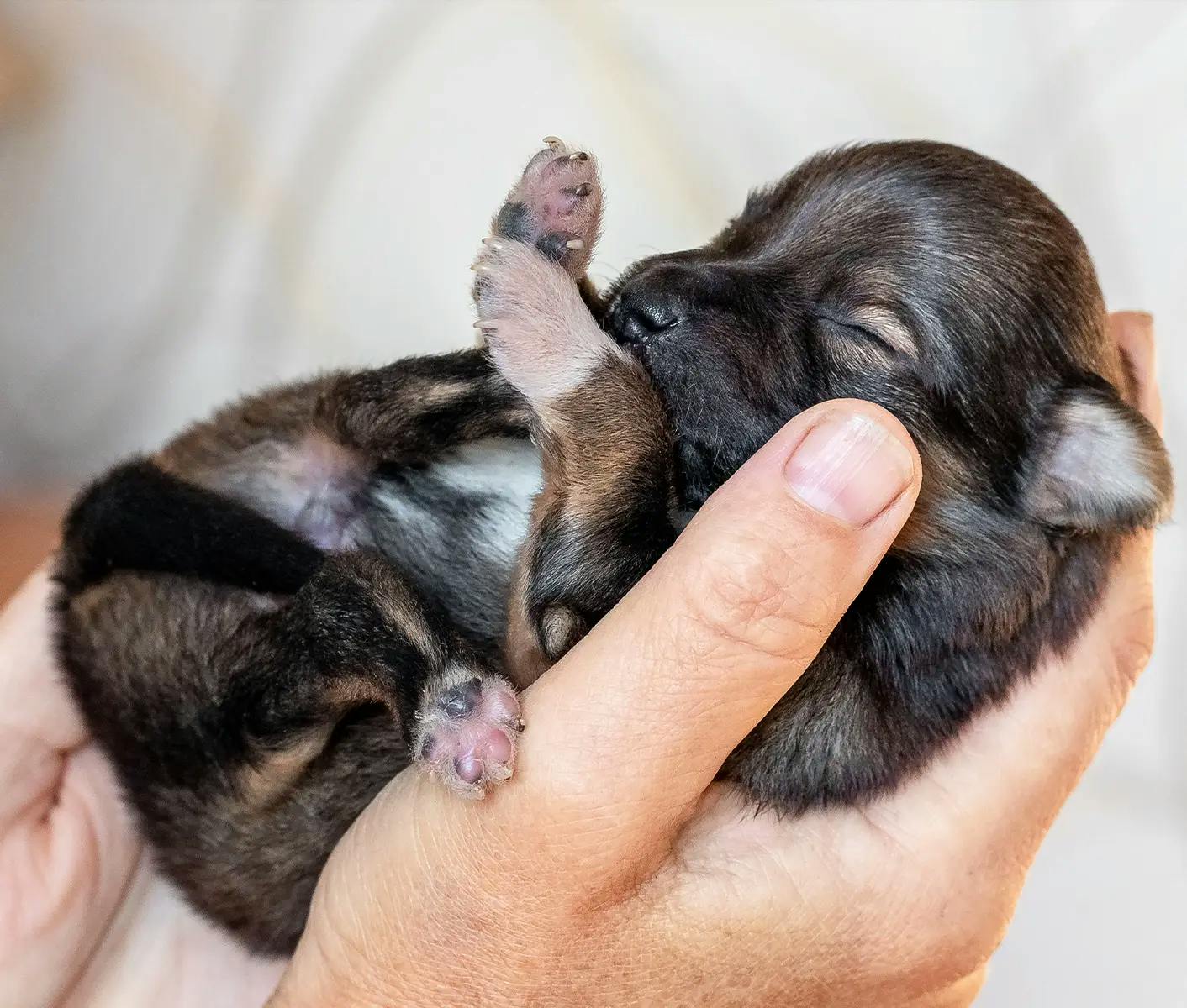
Kitten or Puppy With Umbilical Hernia
If a pup or pups are produced that do have a puppy belly button hernia, the recommendation is to correct them surgically if they don’t close on their own (many do) and at the time of spaying or neutering if they don’t close.
Umbilical hernias are genetic disorders in most breeds and most cases. However, they can easily be corrected surgically if indicated. It is exceptionally rare to need to use mesh or other complex surgical techniques to close the vast majority of umbilical hernias. This single genetic condition should not be a reason to eliminate mildly affected dogs from a breeding program if the dog has other qualities that merit the inclusion in a program.
Other Types of Hernias in Dogs & Cats
There are several other disorders seen in mammals that are similar to an umbilical hernia and may add confusion to the discussion.
Gastroschisis in Puppies and Kittens
Gastroschisis is when a kitten or puppy’s exposed intestines protrude outside abdomen through an opening off to the side of the belly button/umbilicus, but they are not covered by a protective membrane. Because the intestines are not covered by a sac, they can be damaged by exposure to amniotic fluid in utero, which causes inflammation and irritation of the intestine. This can result in complications such as problems with movements of the intestines, scar tissue, and intestinal obstruction. It is also difficult to keep the intestines and other organs sterile, moist, contained, and undamaged during birth and handling shortly after birth while arranging surgical correction.
Omphalocele
Omphalocele occurs when the newborns intestines, liver or other organs protrude outside the abdomen though the umbilicus. Embryologically, as the puppy develops during the first trimester of pregnancy, the intestines get longer and push out from the belly into the umbilical cord. The intestines normally go back into the belly. If this does not happen, an omphalocele occurs. The omphalocele can be small, with only some of the intestines outside of the belly, or it can be large, with many organs outside of the belly.
In this situation, the organs are covered with a thin, transparent sac of peritoneal tissue. There are often other associated birth defects including heart and kidney defects. Additionally, the abdominal cavity may not be large enough to accommodate the organs when replacing them surgically. In humans, it is associated with heart and neural tube defects as well as other genetic syndromes. An omphalocele is worse than gastroschisis – it has more associated anomalies and a higher rate of mortality than gastroschisis.
In humans, there is an increased risk of omphalocele when the mother smokes, drinks alcoholic beverages, takes SSRI antidepressants, is obese, or is a teenager. Since most of our dogs and cats are not smokers or drinkers, are not on antidepressants or are teenagers, we don’t have a good answer for the cause of these disorders.
In humans, these hernias may be detected by ultrasound prior to the birth of the child. In these cases, the baby may have surgery while in utero or immediately after birth. These are not options in dogs and cats.
Kitten or Puppy Born with Intestines Outside of Body
When a kitten or puppy is born with intestines exposed, whether an omphalocele or gastroschisis, immediate surgery is necessary. If the pup or kitten is born at the veterinary hospital, there is a better chance of successful interventional surgery. However, despite the best efforts of the veterinary team, some newborns cannot or should not be saved. Surgery includes protecting the organs while transporting and preparing for surgery, keeping more intestines from pushing out of the abdominal cavity while handling, keeping the intestines sterile, and protected from damage, anesthesia of the newborn pup, enlarging the abdominal wall defect to reposition organs into the abdominal cavity, appropriate suture techniques, post op antibiotics, and post op pain medications. For most born at home, this cannot be accomplished. For some kittens or puppies born by C-section, this can be accomplished with quick thinking veterinary team members, a skilled surgeon, owners willing to put forth the money and effort, no additional genetic disorders, and a lot of luck.
Inguinal Hernias in Dogs and Other Dog Hernias
Other hernias seen in humans and animals include inguinal hernias (in the groin region), diaphragmatic hernias, peritoneal-pericardia diaphragmatic hernias (PPHD), perineal hernias (next to the rectum in the intact male dogs) and traumatic hernias anywhere on the body cavity. Inguinal hernias are second to umbilical hernias in frequency. An open thoracic wall rarely occurs. In this case, the pup can rarely be saved as there is usually inadequate chest wall (ribs and skin) to close. Additionally, surgical intervention is too slow to keep the pup breathing during intervention.
Other midline defects (defects that occur on the front of the body) also include cleft palate, cleft lip, open thoracic wall, open fontanelle, and spina bifida.
Can You Breed a Dog With an Umbilical Hernia?
When it comes to breeding a dog with an umbilical hernia, it depends on several factors.
One is that you don’t breed a female to a male with the same disorder. A second is that it should be surgically corrected prior to the weight of a pregnancy expanding the hernia if the hernia is large. If the hernia is relatively small, it can be corrected at the female’s first c-section if one is planned or expected. A third is IF and only if she brings other great positive traits to a breeding program, including a fit temperament, normal to superior orthopedic clearances, good conformation, and no other serious health conditions considered to be inherited.
In my opinion, we need to rate genetic and congenital disorders based on severity. I rate disorders on a scale of one through three. To me, level one is a minor disorder that is easy to live with or easy to correct. This includes umbilical hernias, distichia (extra eyelashes), entropion (rolled in eyelids), and retained testicle(s).
There are some veterinary experts who recommend avoiding umbilical hernia dog breeding, stating that these dogs when bred will have progressive severity, resulting in gastroschisis and omphalocele. Other veterinary experts do not believe this is the case.
A concern many veterinarians have is the risk of abdominal organ strangulation and/or entrapment if the umbilical hernia is left unmanaged surgically. According to the unpublished literature, this condition is rare and is easily managed if it becomes a concern. The AKC and AVMA allow and encourage the surgical correction of umbilical hernias should this be medically indicated.
If you have questions on cat or dog hernias, call us at 800.786.4751.
LEARN MORE:
Newborn Puppy Care: Managing Neonates and High-Risk Puppies
Improve neonatal survival outcomes when puppies are in trouble. Dr. Greer provides resources to measure and strengthen the health of newborn puppies.


Kitten and Puppy Umbilical Cord Care
What do I need to know when cutting a puppy umbilical cord? Kitten and puppy umbilical cord care is often forgotten. Learn how to properly care for a kitten or newborn puppy umbilical cord to avoid navel infection.
Vet Minute: Umbilical Cord Care for Newborn Puppies
Do puppies umbilical cords get infected? Learn about puppy umbilical cord infection and how to care for a newborn puppy umbilical cord.

Dog Breeding Genetics: Maintaining Good Canine Genetics
What testing should be done before breeding dogs? When breeding dogs, we want to maintain the good genetics and breed away from detrimental issues. Learn about dog genetics and genetic testing for dogs before breeding.

Written by: Marty Greer, DVM
Director of Veterinary Services
Marty Greer, Doctor of Veterinary Medicine, has 40+ years’ experience in veterinary medicine, with special interests in canine reproduction and pediatrics. She received her Doctor of Veterinary Medicine from Iowa State University in 1981. She’s served as Revival’s Director of Veterinary Services since 2019. In 2023, Dr. Greer was named the Westminster Kennel Club Veterinarian of the Year.